
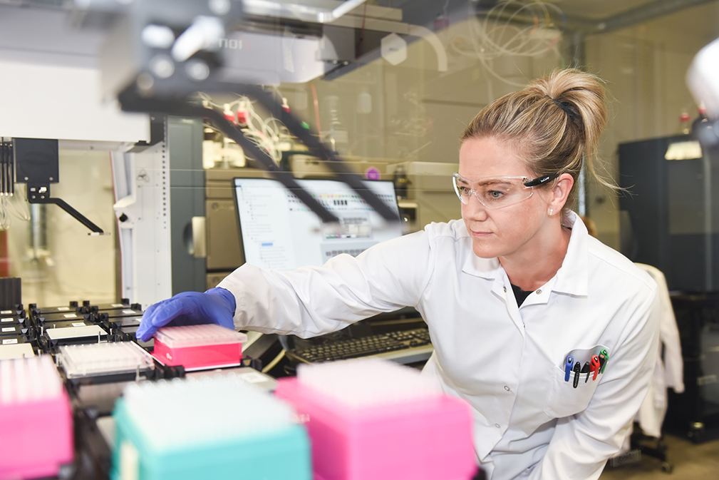
Assessing the ability of a new drug candidate to inhibit major drug-metabolizing enzymes is a standard and essential part of modern drug discovery and development. Because the Cytochrome P450 (CYP) enzyme family plays a central role in the metabolism of most drugs, preclinical drug–drug interaction studies primarily focus on CYP inhibition. Additional drug-metabolizing enzymes, including UDP-glucuronosyltransferases (UGTs), may also be evaluated, particularly when the investigational drug is identified as a substrate of these enzymes.
Inhibition of CYP enzymes is generally classified as either reversible inhibition or time-dependent inhibition (TDI), both of which are critical considerations in drug metabolism and drug–drug interaction studies. Reversible CYP inhibition occurs through rapid association and dissociation between the inhibitor and the enzyme and is commonly divided into three categories: competitive, noncompetitive, and uncompetitive inhibition. Competitive inhibitors bind directly to the enzyme’s active site, whereas noncompetitive and uncompetitive inhibitors bind to allosteric sites located elsewhere on the enzyme.
Time-dependent inhibition (TDI) of CYP enzymes refers to inhibition that increases after preincubation of the enzyme with the inhibitor. TDI is often associated with the formation of reactive intermediates or inhibitory metabolites that cause irreversible or quasi-irreversible enzyme inactivation. In irreversible inhibition, enzyme activity is permanently lost because of covalent binding to the enzyme, while quasi-irreversible inhibition typically involves strong non-covalent interactions. Because CYP enzyme activity can only recover after synthesis of new enzymes, irreversible inhibition may lead to prolonged and clinically significant drug-drug interactions. Therefore, it is recommendable to include time-dependent CYP inhibition screening to the DMPK strategy early on.
Distinguishing between reversible CYP inhibition and irreversible or time-dependent inhibition has therefore become an important aspect of preclinical safety assessment. Although the terms time-dependent inhibition and irreversible inhibition are frequently used interchangeably, they are not identical. TDI specifically describes the observed increase in inhibitory potency following preincubation with an inhibitor. Additional mechanistic studies, including dialysis assays, are typically required to determine whether the inhibition mechanism is reversible, quasi-irreversible, or truly irreversible.
According to the ICH M12 Guideline, the inhibitory potential of an investigational drug—covering both reversible inhibition and time-dependent inhibition—should at a minimum be evaluated against key Cytochrome P450 (CYP) isoforms. These include CYP1A2, CYP2B6, CYP2C8, CYP2C9, CYP2C19, CYP2D6, and CYP3A, which collectively account for the majority of clinically relevant drug metabolism and DDI risk. Additional CYP isoforms or other drug-metabolizing enzymes may also be investigated when scientifically justified. This is particularly important if the investigational compound is identified as a substrate of a specific enzyme or is expected to be co-administered with known substrates of that pathway. To ensure clinical relevance, test compound concentrations should reflect anticipated clinical exposure levels. For orally administered drugs, it is also important to consider intestinal exposure based on the expected maximum clinical oral dose, particularly to assess potential inhibition of intestinal CYP3A-mediated metabolism. This is critical for accurately predicting first-pass drug-drug interactions.
In many cases, achievable test concentrations are limited by the compound’s solubility in the experimental matrix. Therefore, a preliminary kinetic solubility assessment is often used as a practical starting point for defining experimental conditions and optimizing study design in preclinical CYP inhibition assays.
If CYP inhibition is observed, the kinetic parameters describing the inhibition in more detail can be determined in a follow-up study. In the case of reversible inhibition, this involves the determination of the inhibition constant Ki, the equilibrium constant for the formation of an enzyme-inhibitor complex. For time-dependent inhibition, the corresponding parameters are kinact and KI, which define the maximal rate of enzyme inactivation and the inhibitor concentration yielding half maximal inactivation rate. For reversible inhibition, the M12 guideline allows the estimation of Ki based on observed IC50 values using IC50,u/2 as a conservative estimate for Ki.

Experimentally, enzyme inhibition drug-drug interaction studies typically involve incubating an enzyme-specific probe substrate in both the absence and presence of a test compound across a range of concentrations. The effect of the compound is then evaluated by measuring changes in the formation rate of the probe substrate, which serves as a direct indicator of enzyme activity and inhibition potency. This approach is widely used in drug metabolism studies and drug–drug interaction (DDI) risk assessment. To improve throughput and cost efficiency, inhibition assays are often performed using a cocktail approach, where probe substrates for CYP isoenzymes are combined in a single incubation. The resulting metabolites are then quantified simultaneously using LC–MS/MS bioanalytical methods, enabling efficient multiplex analysis of CYP inhibition across several enzymes in one experiment.
The most commonly used enzyme source in CYP inhibition studies is human liver microsomes (HLM). These microsomes contain the major drug-metabolizing CYP enzymes present in vivo and also include other enzyme systems such as UDP-glucuronosyltransferases (UGTs). For CYP-specific studies, isoform-selective probe substrates are available, enabling robust application of cocktail-based CYP inhibition assays.
When selective probe substrates are not available, recombinant enzyme systems may be used as an alternative to isolate enzyme-specific activity. However, subcellular systems such as microsomes may under- or overpredict in vivo drug–drug interaction risk, particularly for compounds that undergo significant non-CYP-mediated metabolism not captured in these systems. In such cases, primary human hepatocytes, which retain full metabolic capacity and membrane transport activity, may provide a more physiologically relevant model for predicting in vivo CYP inhibition and overall DDI risk.
A key principle in drug metabolism research is the free drug hypothesis, which states that only the unbound (free) fraction of a drug is pharmacologically active. This concept is equally critical for enzyme inhibition, as only unbound drug concentrations can interact with and inhibit metabolizing enzymes. The importance of using unbound drug concentrations is also emphasized in the ICH M12 Guideline, which requires incorporating unbound plasma and unbound incubation concentrations when assessing DDI potential.
The unbound fraction in incubation systems (fu,inc) can be determined experimentally using methods such as rapid equilibrium dialysis (RED) or ultrafiltration, or estimated computationally using predictive models based on physicochemical properties such as lipophilicity, charge, and protein binding affinity. Accurate estimation of fu,inc is essential for translating in vitro inhibition data into clinically relevant predictions of drug–drug interaction risk.
Want to discuss your DDI risk assessment strategy or learn how CYP inhibition studies can be integrated into your broader DMPK workflow? Our experts are here to help — reach out to us to continue the conversation.